Hint Novel Therapies
Naming
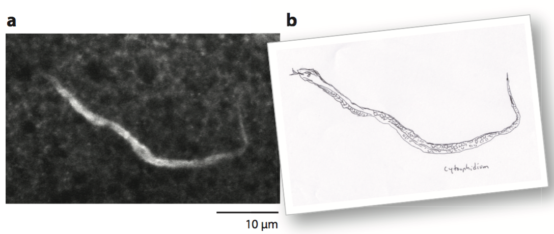
The cytoophidium: a snake in the cell. Liu (2010) refered to these subcellular snake-like structures as cytoophidia (Greek: cyto-, meaning cell, and ophidia, meaning serpents) or cytoophidium (singular). (a) A snake-like structure observed in a Drosophila oocyte. This was one of the first images of cytoophidia obtained by antibody cross-reaction. Adapted with permission from . (b) A drawing of a snake mimicking the image in panel a.
Cytoophidia of IMPDH and CTPS
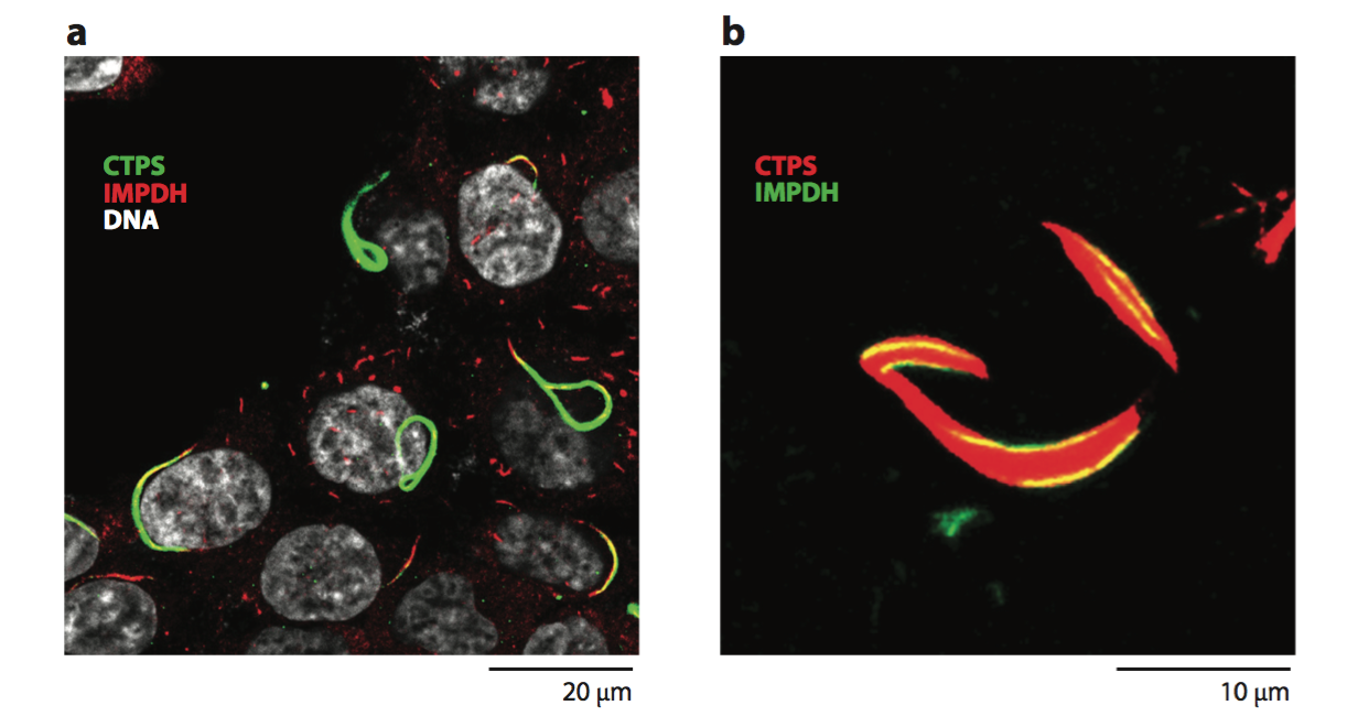
(a) CTPS (green) and IMPDH (red) can form independent cytoophidia. CTPS and IMPDH cytoophidia sometimes overlap (yellow). (b) Thin IMPDH cytoophidia (green) attach to the surfaces of thick CTPS cytoophidia (red). CTPS was overexpressed in both panels. Panel a courtesy of Chia Chun Chang and Li-Ying Sung. Panel b modified from Chang et al. (2015).
Assembly
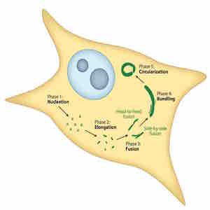
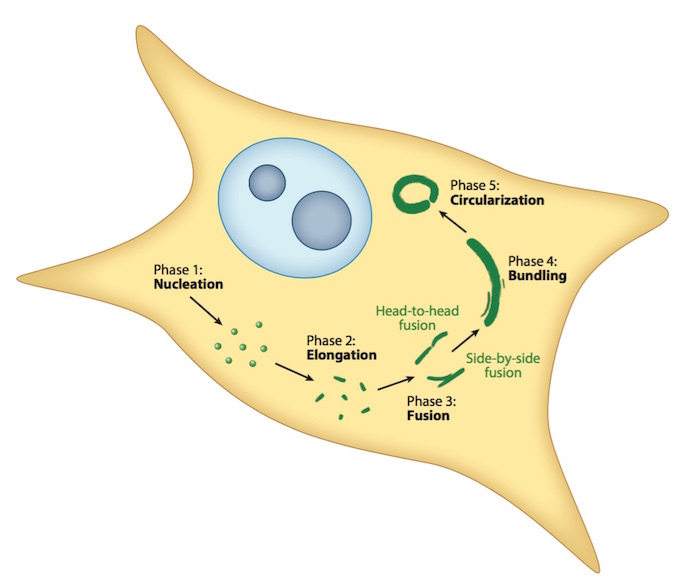
The five phases of cytoophidium assembly: (1) nucleation, (2) elongation, (3) fusion, (4) bundling, and (5) circularization. Modified from Gou et al. (2014).
Interplay between Myc and Cytoophidia
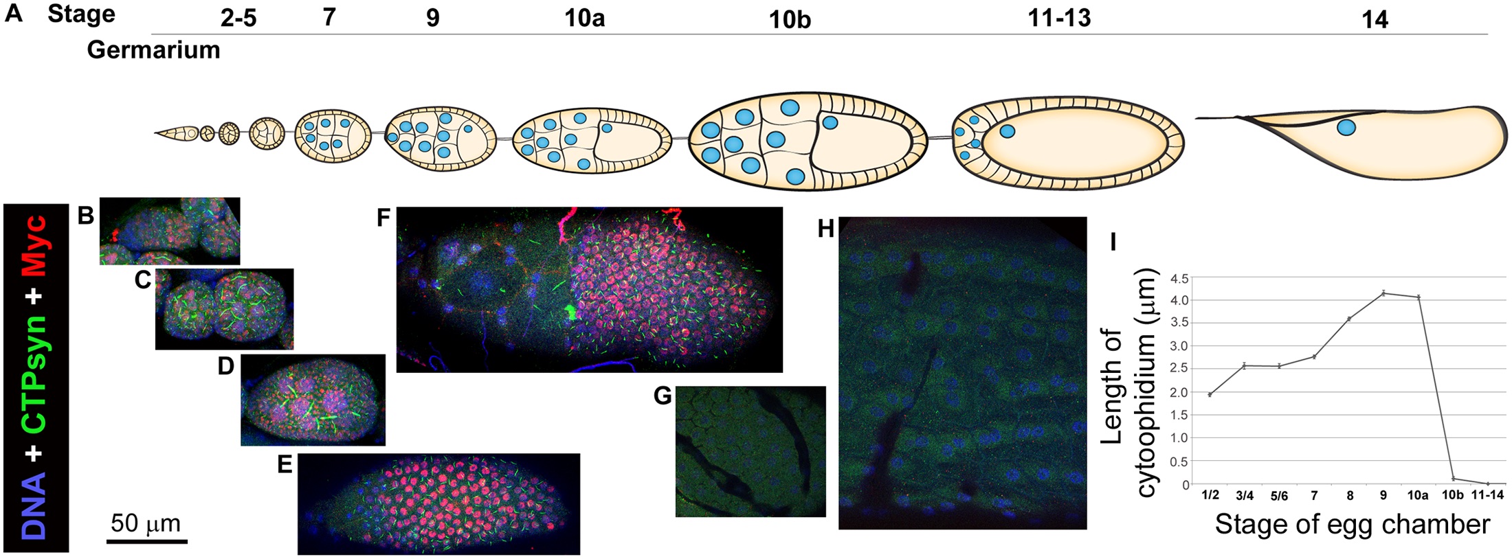
Cytoophidium formation correlates with Myc expression in Drosophila follicle cells.
|
1. Liu JL. (2016). The cytoophidium and its kind: Filamentation and compartmentation of metabolic enzymes. Annual Review of Cell and Developmental Biology 32:349-72. [HTML] 2. Aughey GN, Grice SJ and Liu JL. (2016). The interplay between MYC and CTP synthase in Drosophila. PLOS Genetics 12(2):e1005867. [HTML] 3. Ghosh S, Tibbit C and Liu JL. (2016). Effective knockdown of Drosophila long non-coding RNAs by CRISPR interference. Nucleic Acids Research 44 [Epub on 4 Feb 2016]. [HTML][OXfCRISPR] 4. Bassett AR, Tibbit C, Ponting CP and Liu JL. (2013). Highly efficient targeted mutagenesis of Drosophila with the CRISPR/Cas9 system. Cell Reports 4(1):220-8. [HTML][OXfCRISPR][Cell Reports Best of 2013] 5. Liu JL. (2010). Intracellular compartmentation of CTP synthase in Drosophila. Journal of Genetics and Genomics 37(5):281-96.[HTML][NATURE | News Feature] 6. Liu JL, Murphy C, Buszczak M, Clatterbuck S, Goodman R and Gall JG. (2006). The Drosophila melanogaster Cajal body. Journal of Cell Biology 172(6):875-84.[HTML] |
Waiting for talents like you guys to kick off.
